

How do I digitize a radiograph?
Digital Radiography
As in other NDT methods, the introduction of microprocessors and computers has brought about significant changes to radiographic examination. Chapter 17 describes a number of systems such as computed-tomography and radioscopy which are made possible by newly developed technology for the rapid digital processing of vast quantities of data.
But as this chapter (16) shows, computer technology has also entered the field of conventional image forming radiography, as applied in industry. The driving force was the medical world where digital radiography already earned its credits and has become standard technology. Along with a few other companies, Waygate Technologies has developed digital systems with a wide range of computer-aided NDT applications. Partly digital radiography does replace conventional film and to some extend also permit new applications.
The following three main methods can be identified:
- Digitization of conventional flexible X-ray films for the purpose of archiving and/or image enhancement (manipulation)
- Digital radiography by means of phosphor coated semi-flexible imaging plates and computer processing, so-called “Computed Radiography” or CR
- Digital radiography e.g. with rigid flat-panel detectors and instant computer processing, referred to as “Digital Radiography” or DR, sometimes referred to as “Direct Radiography”.
Each method has differing strengths, advantages and limitations that should be evaluated in terms of specific application, inspection requirements and economics: capital, human investment and production (number of exposures in a certain time).
The major merits of digital radiography compared to conventional film are:
- Shorter exposure time and thus potentially safer
- Faster processing
- No chemicals, thus no environmental pollution
- No consumables, thus low operational costs
- Plates and panels can be used repeatedly
- A very wide dynamic exposure range/latitude thus fewer retakes
On the other hand image resolution of even the most optimized digital method is (still) less than can be achieved with finest grain film. A few other limitations are also explained in this chapter.
How do I digitize a radiograph?
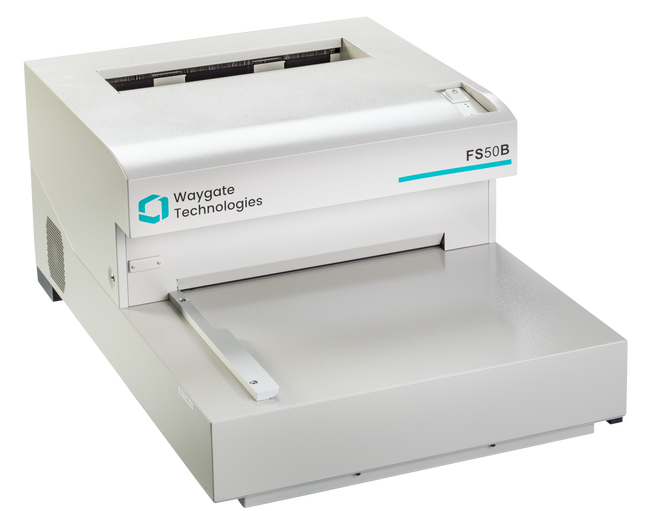
Storing and archiving of chemically developed X-ray films not only demands special storage conditions, see section 10.7, but also takes up quite a bit of space. Digitisation of these films provides an excellent alternative that also prevents degrading. Special equipment has been developed for this purpose.
Current digitisation equipment actually consists of a fast computer-controlled scanner that scans the film spotwise in a linear pattern, identical to the formation of a TV-image, measuring densities while digitising and storing the results. The laser beam spot can be as small as 50 μm (micron, equivalent to one thousand’s of a millimetre) diameter, but the equipment can be adjusted for a coarser scan, for example 500 micron, and therefore shorter scanning times.
The values measured are compared with a calibrated density scale and processed digitally. Density variations between 0.05 up to 4.7 can be measured and digitised with e.g. 12 bit density steps (4096 grey levels), equivalent to almost 0.001. Films with a maximum width of 350 mm can be digitised in a single run. Even for the smallest pixel size of 50 μm, approximately 4 mm of film can be scanned per second, so for the largest standard film size (350 x 430 mm) this process would take approximately 2 minutes.
Scanners exist without length limitation of film, and adapters are available for digitisation of roll films.
Apart from the greatly reduced storage space and (almost) deterioration-free archiving, digitising also makes it possible to (re)analyse the film’s images on a computer screen (see figure 18-16), with the possibility of electronic image manipulation.
Thus defect indication details not discernible on the original film using a viewing screen can be made visible.
Because scanners vary widely in resolution, dynamic range, and ability to scan dense films, evaluation is required to ensure that adequate scanning fidelity is achieved. Depending on the selected resolution many Megabytes are needed to store a single film. Archiving usually is done on a mass storage facility e.g.: CD-ROM, DVD etc.
For use in laboratory environments only, high resolution film digitisation systems exist applying a scanning spot size of 10 μm. This enables detailed analysis of particular film areas.
What types of material are used for radiographic intensifying screens?