What is an anode?
The target is generally made of tungsten. Not only because it has a high atomic number, but also because of its high melting point (approx. 3400˚C). It is essential to use a material with a high melting point because of the substantial amount of heat dissipated as the electron-“bombardment” is concentrated (focused) on a very small surface. Only a part (approx. 0.1 % at 30 keV; 1 % at 200 keV; 40 % at 30 to 40 MeV) of the kinetic energy of the electrons is converted into X-radiation; the remainder is trans- formed into heat.
Cooling the anode: The heat which accompanies the production of X-radiation is quite considerable, so that the anode has to be cooled. This can be done in a variety of ways : 1. by natural radiation 2. by convection 3. by forced circulation of liquid or gas 4. by conduction
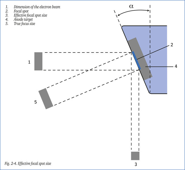
The focal spot: The area of the target which is struck by the electrons, see figure 2-4, is called the focal spot or “the focus”. It is essential that this area is sufficiently large to avoid local overheating, which might damage the anode. From the radiographic point of view, however, the focus has to be as small as possible in order to achieve maximum sharpness in the radiographic image. This “focal loading” is expressed in Joule/mm2. A tungsten target can take a maximum loading of 200 Joule/mm2. A higher loading might damage the anode.
Effective focal spot size: The projections of the focal spot on a surface perpendicular to the axis of the beam of X- rays is termed the “ effective focal spot size” or “ focus size”, see figure 2-4. The effective focus size is one of the parameters in radiography, see section 11-1. The effective focus size has to be as small as possible in order to achieve maximum sharp- ness in the radiographic image. The dimensions of the focus are governed by: 1. The size of the focal spot, and 2. The value of angle α, see the figure
It should be noted that when in radiography we speak of the “size of the focus” without spe- cifying this more exactly, it is normally the effective focal spot size which is meant. Conventional X-ray tubes have effective focal spot sizes in the range 4 x 4 mm to 1 x 1 mm. There are fine-focus tubes with focal spots from 0.5 x 0.5 mm ad micro-focus tubes down to 50 μm diameter or even less. The effective focal spot size is determined in accordance with the procedure described in EN 12543. For a practical alternative method see an later section.