

What is direct radiography (DR)?
Digital radiography is also known as direct radiography, for short DR. With DR-technology, there is an immediate conversion of radiation intensity into digital image information. Exposure and image formation happen simultaneously, allowing near real-time image
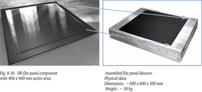
capture, with the image/radiograph available for review only seconds after the exposure. This almost instant image formation is the reason that DR is considered the only genuine method of digital radiography. Some of the devices even provide a true real-time (radioscopic) mode with display rates up to 30 images per second. For DR, flat panel detectors in a variety of sizes are used, up to approximately 400 x 400 mm (maximum in 2006) as shown in figure 8-16, that convert incident radiation intensity into proportional and digitized electronic signals. These digital signals can, by means of a computer and screen (workstation), without intermediate steps, be presented as a coherent radiographic image. A cable typically links
the detector to this workstation from which the panel is controlled as well. There are different types and suppliers of DR-flat panel systems. A variety of flat panel systems exist with a wide range of pixel sizes and resolutions. The more and smaller the pixels the higher the potential resolution of the system. As sensor materials amorphous silicon and amorphous selenium are in use. As sensors CCD’s (Charge Coupled Devices) and CMOS (Complimentary Metal Oxide
Semiconductor) are applied.
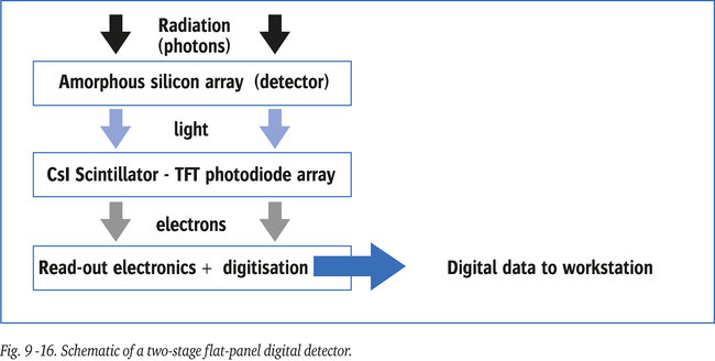
The most common high resolution flat panels use amorphous silicon technology. This material converts incident radiation into light. The conversion is proportional to the radiation dose. This light in turn is converted from light into a proportional electric signal by a scintillator made of e.g. structured Cesium Iodide (CsI) photodiodes and integrated thin film transistors (TFT’s). Each picture element (pixel) contributes to the radiographic image formed on the screen of the workstation. Each element is square in effective area, with pixel pitch typically ranging
from 50 to 400 micron. The smaller the pixels the better the resolution. Development is in progress to make sensor elements/pixels smaller. Depending on overall active area and detector pixel pitch, a panel consists of up to several millions of such elements/pixels. Figure 9-16 shows the different active layers of a flat panel detector which are deposited on a glass substrate with a graphite cover on top.
In practice DR has proven to be an excellent tool for the NDT-industry, however some
limitations apply as well:
- Flat panel detectors can be used continuously for years in mass production processes, to some extent however their lifetime is limited by the accumulated radiation dose. The ultimate lifetime is determined by a combination of total dose, the dose rate and radiation energy. The plates are less tolerant for high than for low energy radiation, hence extreme high energies should be avoided. Thus the ultimate lifetime is dependent on its application.
- With the millions of pixels it is “normal” that over time that a few pixels become less responsive, similar to pixels of flat panels as used with (notebook)computers. Usually the un-acceptable number and pattern of dead pixels is specified by the manufacturer. Fortunately, in cases a small area of the panel is out of order, an experienced interpreter of DR-images is able to differentiate (by pattern recognition and known position on the panel) real component defects from less responsive pixels.
- Flat plate detectors are also subject to some memory effect, in jargon called “ghosting”. This is due to hysteresis of the scintillation layer after exposure. The image slowly fades away, particularly in case of high energy levels exceeding a few hundred kV. This hysteresis causes a certain dead time of the system, from seconds to minutes depending on the radiation energy, during which the plate cannot be re-used.