What is fluoroscopy and how are image intensifiers used?
Fluoroscopy, also known as radioscopy, is a technique whereby “real-time” detection of defects is achieved by the use of specialized fluorescent screen technology. At present, there are many alternatives to photographic film for making an X-ray image visible. In addition to the CR and DR techniques described before (chapter 16), a wide range of real-time image forming techniques using display monitors are available. These systems are considered also to belong in the DR category.
It can generally be said that the image quality of conventional X-ray film is superior to either digital direct radiography (DR) or computer-aided film radiography (CR) techniques. Thus these new techniques cannot be considered acceptable alternatives at all times. However, when the installation is adjusted to optimal refinement for a single application, for example weld inspection in a pipe mill, a film equivalent image quality can be obtained, which would only just comply with the requirements. This would e.g. demand the use of a
micro-focus tube, as seen in this section.
Stationary real-time installations
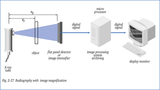
Display monitor systems, as illustrated in figure 2-17, are almost exclusively used in stationary set-ups for production line testing of varying types of objects, in particular in metal-casting plants, pipe mills and component assembly industries. Sometimes real-time systems are utilized in the food industry to check for instance for the
presence of glass fragments or other foreign objects. Being part of a production line and due to the necessary radiation safety provisions (such as cabins) these systems can be very expensive. The display monitors are located at a safe distance.
The choice of a radiographic system to be used depends on a number of factors:
- Hardness of the radiation required and proper detector.
- Resolution or detail discernibility required. The type of defects to be detected in mass-production is normally known.
- Magnification factor required when it concerns small defects.
- Image dynamics (density range) with regard to object thickness range.
- Image contrast required facilitating ease of defect detection. Sometimes this can be “automated” when it concerns common defects.
- Time restraints, number of objects to be examined per unit of time.
- Budget.
- Space available.
- Installation and specimen dimensions.
A number of these factors also influence the choice of detector system.
Some of the options are:
- Phosphorescent screen (afterglow) with TV camera and display monitor (CCTV) at a remote (safe) location
- Fluorescent screen (instant image) with CCTV-system at a safe location
- X-ray image intensifier with conversion screen, in combination with a CCTV-system
- CCD-camera as a substitute for the relatively slow conversion screen
- Photo array detector, minimal size per diode approx. 0.25 mm to inspect slowly moving objects (airport luggage checks)
- Flat panel detector, FPD, consisting of millions of light-sensitive pixels. The light pulses can be converted in different ways into electric pulses, which in turn can be processed digitally.
Although the image intensifier is still most commonly used, the flat panel detector is becoming more and more attractive. Flat panel detectors provide various pixel sizes with extensive image dynamics (a very wide density range, far greater than is possible with film).
Since the signals received by the computer are digital, the screen image can be optimized for interpretation (contrast, brightness, sharpness, magnification, filtering, noise suppression) and subsequently stored. These advanced systems also offer the possibility of comparing the image obtained with a reference image and of automatic defect interpretation.
Selection of the most suitable (expensive) system is made even more difficult because of the rapid development in sensor- and electronic technology.
Fluoroscopy, imageintensifiers and-magnifiers are more elaborately described in the booklets: Die “Röntgenprüfung” and its translation “The X-ray inspection”.
Portable real-time equipment
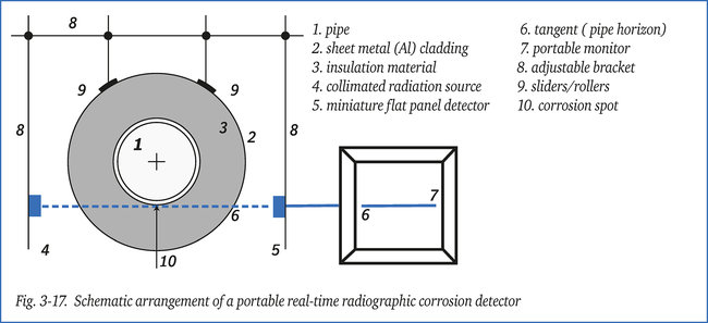
A portable version of real-time equipment is used to detect external corrosion under thermal insulation. It is generally very difficult to detect corrosion on piping with insulation still in place, whereas removing and re-installing the insulation is a costly and time consuming operation. Sometimes the likely presence of corrosion is indicated by moisture/water detected in the insulation, see in a later section. External corrosion in a low-alloy steel pipe becomes apparent by local swelling of the pipe surface as a result of volume increase of the corrosion layer. Figure 3-17 illustrates a system by which the swelling and even severe pitting can be detected. On one side is a strongly collimated source or X-ray tube that must be aligned in such a way as to direct a narrow beam of radiation along the tangent of the pipe towards a flat panel detector behind.
This way an image is obtained of the pipe “horizon” with possible presence of corrosion (swelling or pitting). The image is presented real-time on a portable monitor. The battery-powered equipment uses soft radiation of low intensity, so that it can manually be moved
along the pipe. The system can also be used to locate welds under insulation.
Radiographic Exposure Adjustment in Diverse Wall Thicknesses