How to increase the maximum Scan Volume in Computed Tomography?
In this article:
- Overcoming Detector Size Limitations in CT: Traditional CT systems are limited by detector dimensions and beam cone size, restricting the maximum scan volume and voxel resolution for large objects.
- Virtual|detector Expands Scan Width: By shifting the detector during acquisition, the system captures multiple image segments that are stitched together, enabling scans up to 1.5 meters wide without compromising image quality.
- Multi|scan Increases Vertical Coverage: This technique performs sequential scans at different heights, automatically aligning and merging them to extend the scan volume vertically—ideal for tall components.
- X-offset|scan for Oversized Samples: When objects exceed the system’s width capacity, off-center rotation allows partial projections to be reconstructed into full volumes, maximizing usable scan area.
- Smart Cabinet Design Enhances Accessibility: Features like full-height doors and crane support simplify loading large samples, making the most of available space and improving operator ergonomics.
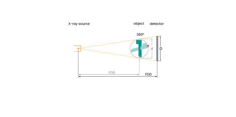
In the early days of industrial CT, the detector size was the limit for the size of the volume that could be scanned without disturbing reconstruction artifacts. The object under examination had to remain within the beam cone. The focus-detector limit is limited, which in turn restricts the magnification and the achievable voxel size. Various means can be used to overcome this limitation.
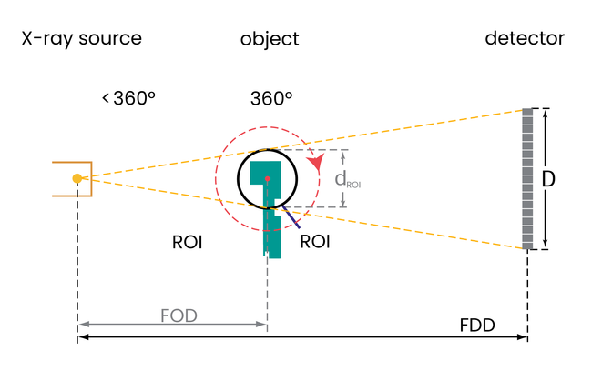
ROI|scan
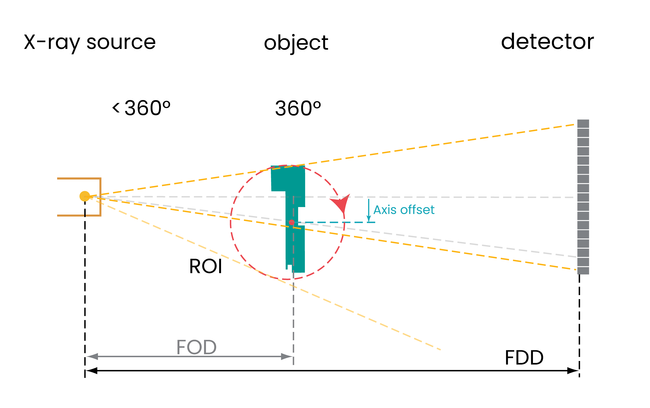
Region of interest scans focus on a part of a larger object that can be captured without loss of quality during reconstruction. Only the part of the object that is in the beam cone during the entire scan is reconstructed, although the object may be much larger.
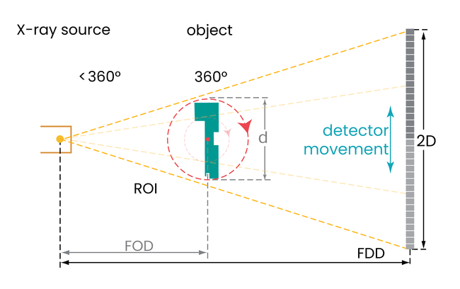
Virtual|detector
The size of the detector is practically increased by moving the detector. For example, the detector moves to the right to capture the right half of a larger image and then to the left to capture the left half of the image. With large devices, the detector can be moved in three, four or five places to make the image even wider. Each projection therefore consists of two or more parts, which are precisely assembled by the software before reconstruction. Such a projection can be 1.5 meters or 60 inches wide.
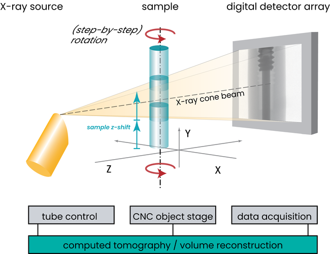
Multi|scan
While Virtual|detector increases the width of the scan volume, the multiple scan function increases the height. The system takes multiple scans to cover the desired height of the object. Between scans, the sample is moved precisely so that the individual volumes fit together when automatically stitched in the reconstruction software. This is only limited by the height of the housing and the travel of the vertical axis.
X-offset|scan
Some machines are too small for a virtual detector or the sample is still too wide. In this case, the sample is rotated off-center during scanning. Only a little more than half of the sample is captured on the projections. Properly processed, the reconstructed volume is almost as good as a Virtual|Detector scan.
Optimized Space in the X-ray system cabinet
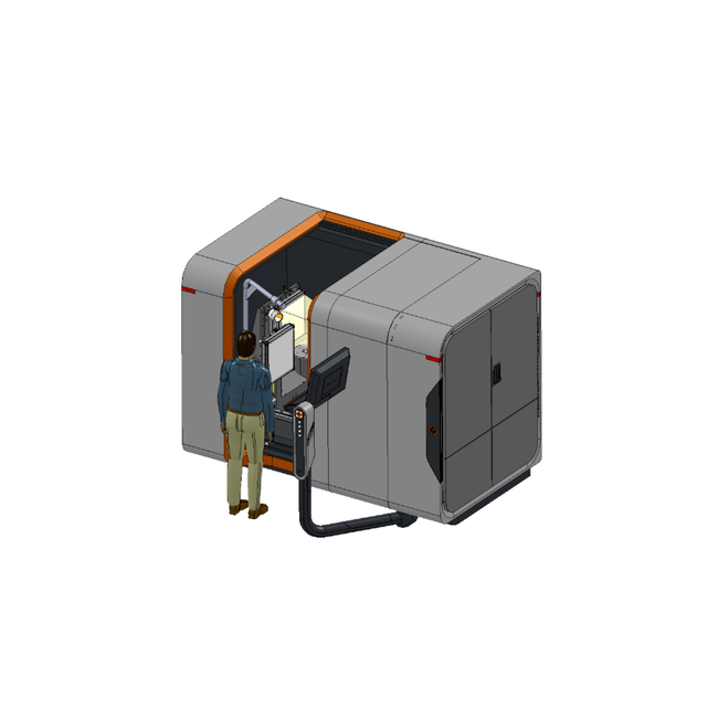
Of course, the sample has to fit through the door and can be difficult to lift. A full-height door that also allows the top of the cabinet to be opened, combined with a crane, therefore ensures easy access for staff. A well-thought-out arrangement can make the interior of the cabinet appear larger than the space it takes up in the laboratory.
