Principles of CT Operation: How does a microfocus X-ray tube work?
In this article:
- Microfocus X-ray Tubes Enable High-Resolution CT Imaging: These tubes generate X-rays from a tiny focal spot—just a few microns wide—allowing for sharp, micrometer-level imaging essential in industrial computed tomography (CT) applications
- Electron Beam Generation and Acceleration: Electrons are emitted from a heated filament in a vacuum and accelerated toward a tungsten target using a high-voltage potential (UACC), where they generate X-rays upon impact
- Magnetic Lenses Focus the Beam: A magnetic lens system narrows the electron beam to a precise focal point, improving image clarity and enabling detailed inspection of small or dense components
- Grid-Controlled Beam Intensity: The Wehnelt electrode (or grid) regulates the electron beam current via bias voltage (UG), offering precise control over X-ray intensity and exposure
- Nanofocus Tubes Push Resolution Boundaries: Advanced transmission-style nanofocus tubes use multiple electron lenses to achieve resolutions down to 200 nanometers, supporting cutting-edge applications in electronics, aerospace, and materials science
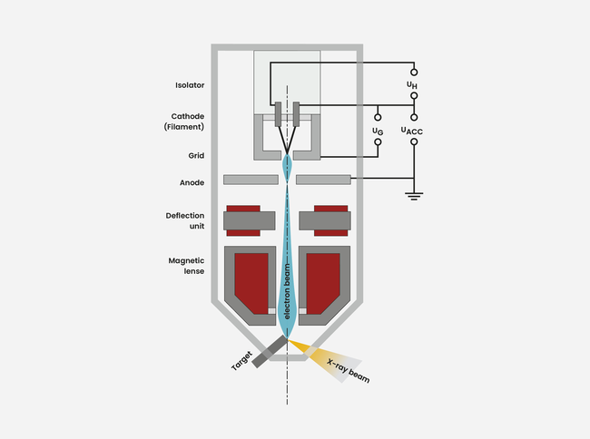
In an evacuated tube electrons are emitted from a heated filament and are accelerated towards the anode by the potential difference UACC. Electrons enter through a hole in the anode into a magnetic lens which focuses the electron beam to a small spot of a few microns in diameter on the massive tungsten target (directional tube).
In the tungsten the electronis are abruptly decelerated whereby X-rays are generated. The focal spot represents a very small X-ray source which enables sharpest imaging with micrometer resolution. Latest nanofocus tubes (transmission tube) achieve a detail detectability down to 200 nanometers (0.2 microns) by using multiple electron lenses. The electron beam current is controlled by the bias voltage UG via the Wehnelt electrode („grid“).

